X-ray Diagnosis
About the Department
The X-ray Diagnostics Department is equipped with modern equipment, with the help the staff of the Department provides qualified specialized diagnostics to patients.
Our medical staff is constantly improving their qualification, undergoing various specializations on topical issues of radiation diagnostics both in Kazakhstan and in countries near and far abroad (Moscow, St. Petersburg, Austria, Prague, Israel, Germany, etc.).
The Department performs such examinations as:
- X-ray diagnostics of organs and systems (scopy and graphy);
- specialized X-ray examinations of organs and systems with contrasting
- digital mammography;
- breast tomosynthesis;
- spectral contrast mammography;
- ductography of mammary ducts of mammary glands;
- osteodensitometry;
- 3D CT of sinuses and jaw, orthopantomography, dental examinations.
All kinds of routine radiographic examinations as well as special radiographic and fluoroscopic examinations are performed in X-ray diagnostic rooms using X-ray machines (manufactured in Germany, USA):
- bone and joint system
- chest organs
- abdominal cavity, etc.
- multiple view fluoroscopy of the esophagus and stomach
- fluoroscopy of the gastrointestinal tract
- bowel passage
- cholangiography
- fistulography
- hysterosalpingography
- cystography
- intravenous excretory urography
- dental examinations (dento-mandibular pathology)
The theory of contrast-enhanced spectral mammography (CESM) is based on the success of dynamic contrast-enhanced MRI of the breast, which is currently the most sensitive of all diagnostic techniques. CESM and MRI with DCU are currently being compared as both techniques are sensitive to vascularization, detect tumor masses, size, shape, and neoangiogenesis. 3D mammography (tomosynthesis) allows:
- to distinguish areas formed by overlapping anatomical structures from apparent nodules
- to detect localized heavy tissue remodeling prior to nodule formation
- microcalcifications, accumulation
- to avoid false-positive and false-negative judgments about the presence of changes on the background of expressed glandular tissue, especially in reproductive age
- to reveal suspicious areas for recurrence of tumor masses
- to observe scar tissue after the surgical period
- to reduce the number of targeted scans and cases of short (3-6 months) dynamic control, thereby reducing the overall radiation burden on the patient
- to reduce the need to use other methods of radiation diagnostics
- to reduce the number of diagnostic puncture biopsies
- Determination of bone mineral density (BMD) of lumbar spine
- Determination of BMD of proximal parts of both femoral bones
- Determination of BMD of the proximal part of the right femur.
- Determination of BMD of the proximal part of the left femur.
Panoramic orthopantomography (panoramic dental image) allows to visualize the condition of the teeth and periapical tissues as accurately as possible, which is necessary for the dentist to determine treatment tactics.
A panoramic dental image allows to obtain an image of the teeth of both jaws and assess their condition using a digital system:
- to detect hidden caries or its recurrence
- to seal failure of fillings
- to determine the condition of filled canals
- to diagnose the condition of bone tissue
Doctors of the Department
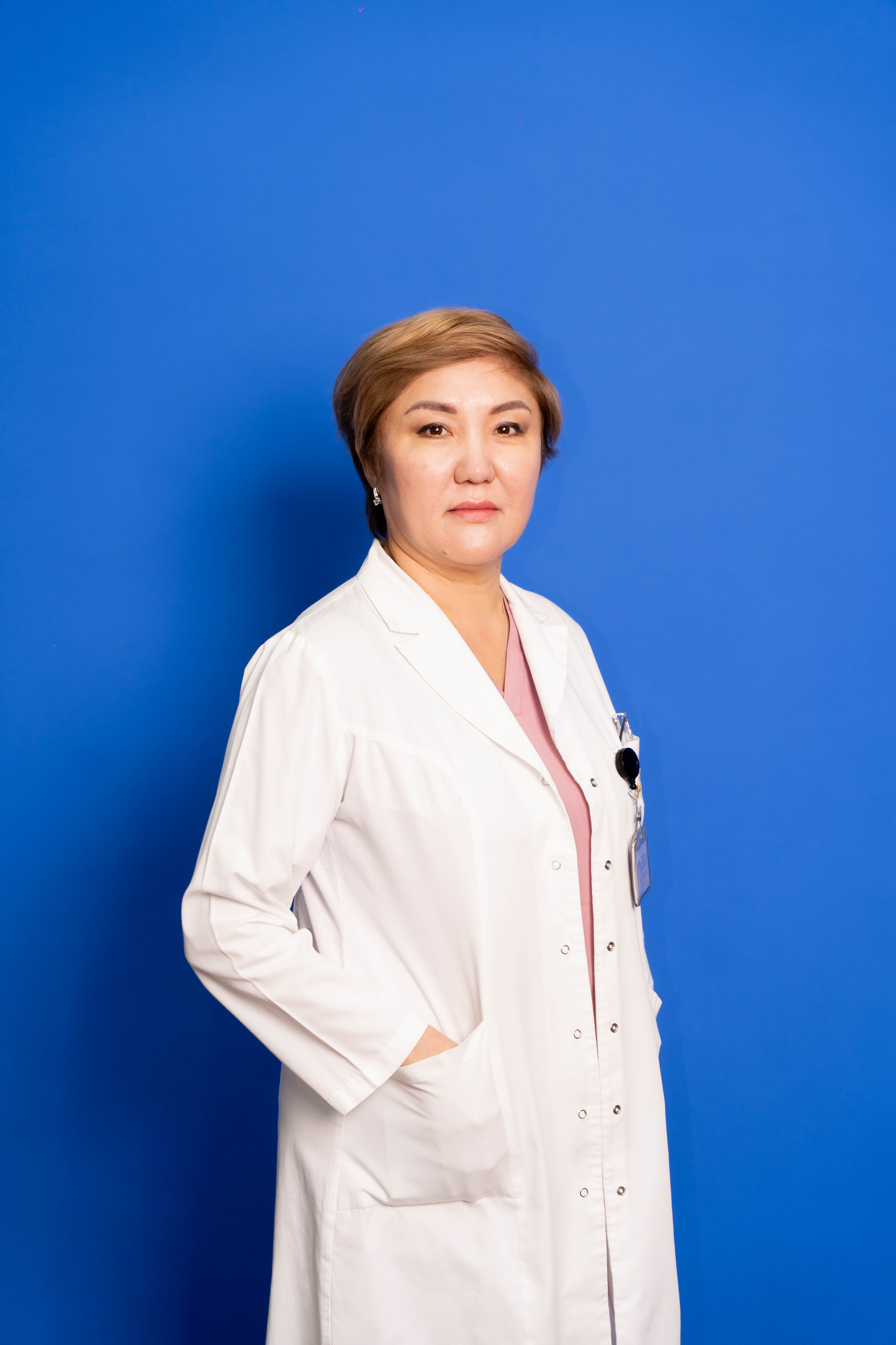 |
Kyzylgul Smailova Chief of the Department The highest category Work experience: 26 years |
|
|
Mahatova Nurzada Iskakovna Radiologist The highest category Work experience: 21 years |
 |
Dinara Khamitova Radiologist The highest category Work experience: 20 years |
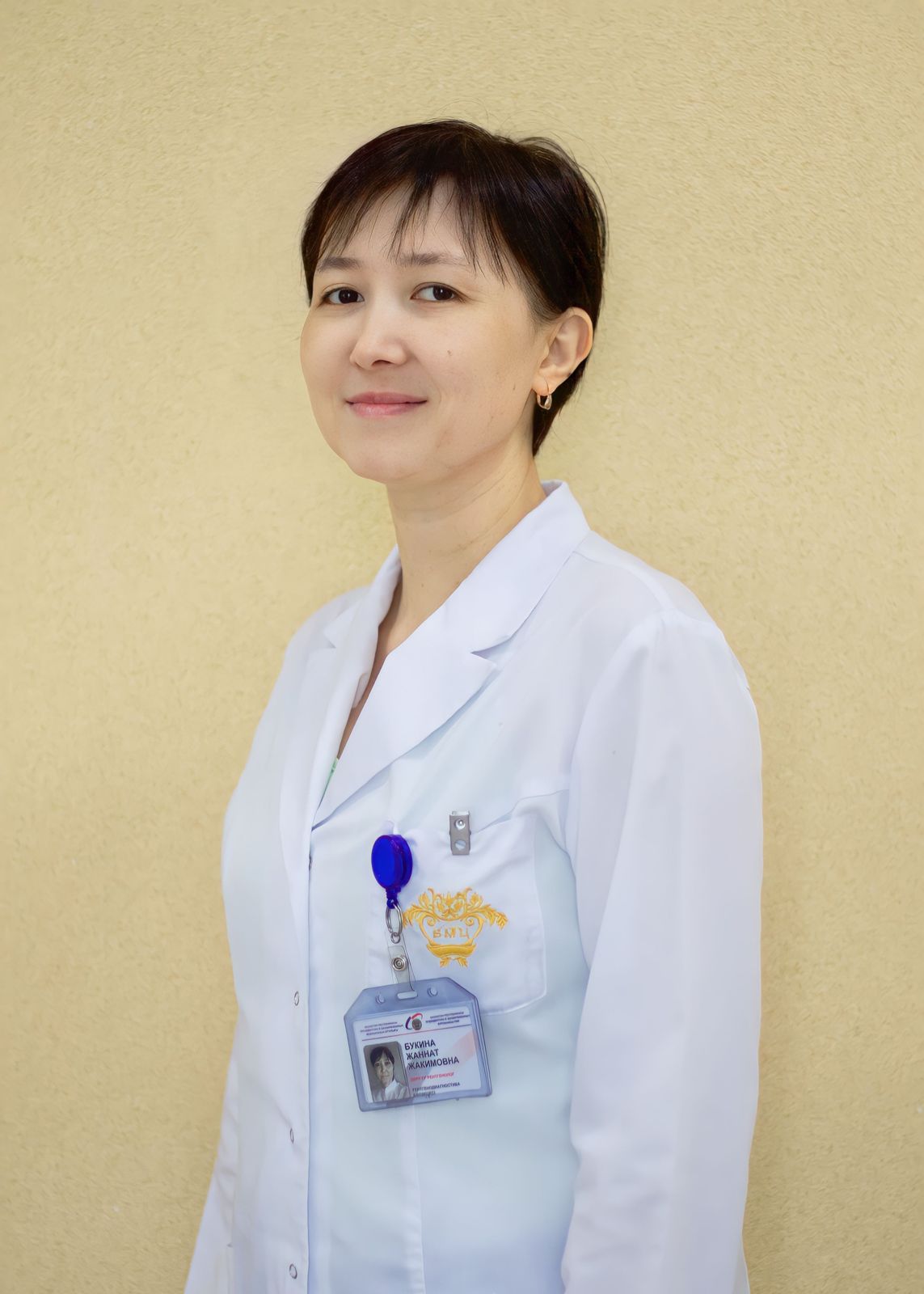 |
Zhannat Bukina Radiologist The highest category Work experience: 20 years |
 |
Sandugash Smailova Radiologist Work experience: 16 years Master of Medicine |
|
|
Miltikbayeva Gulzat Manatbekovna Radiologist The highest category Work experience: 15 years |
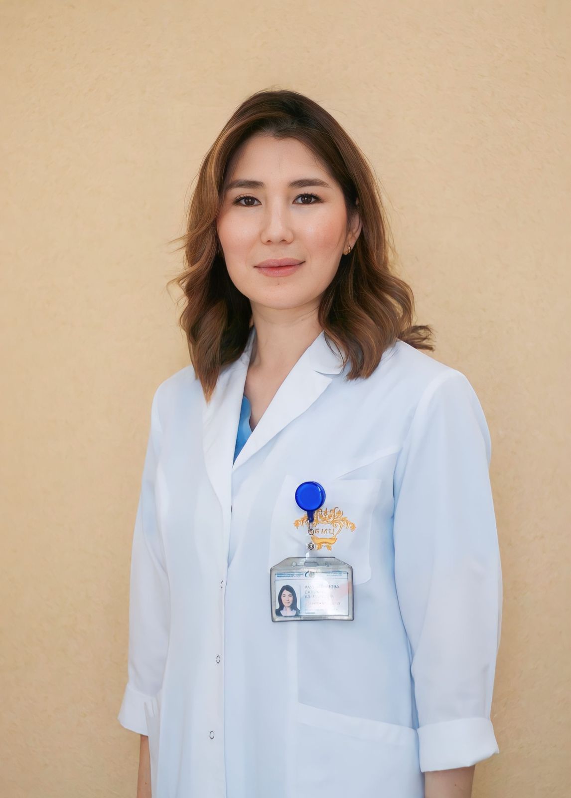 |
Saltanat Rakhmankulova Radiologist The highest category Work experience: 14 years |
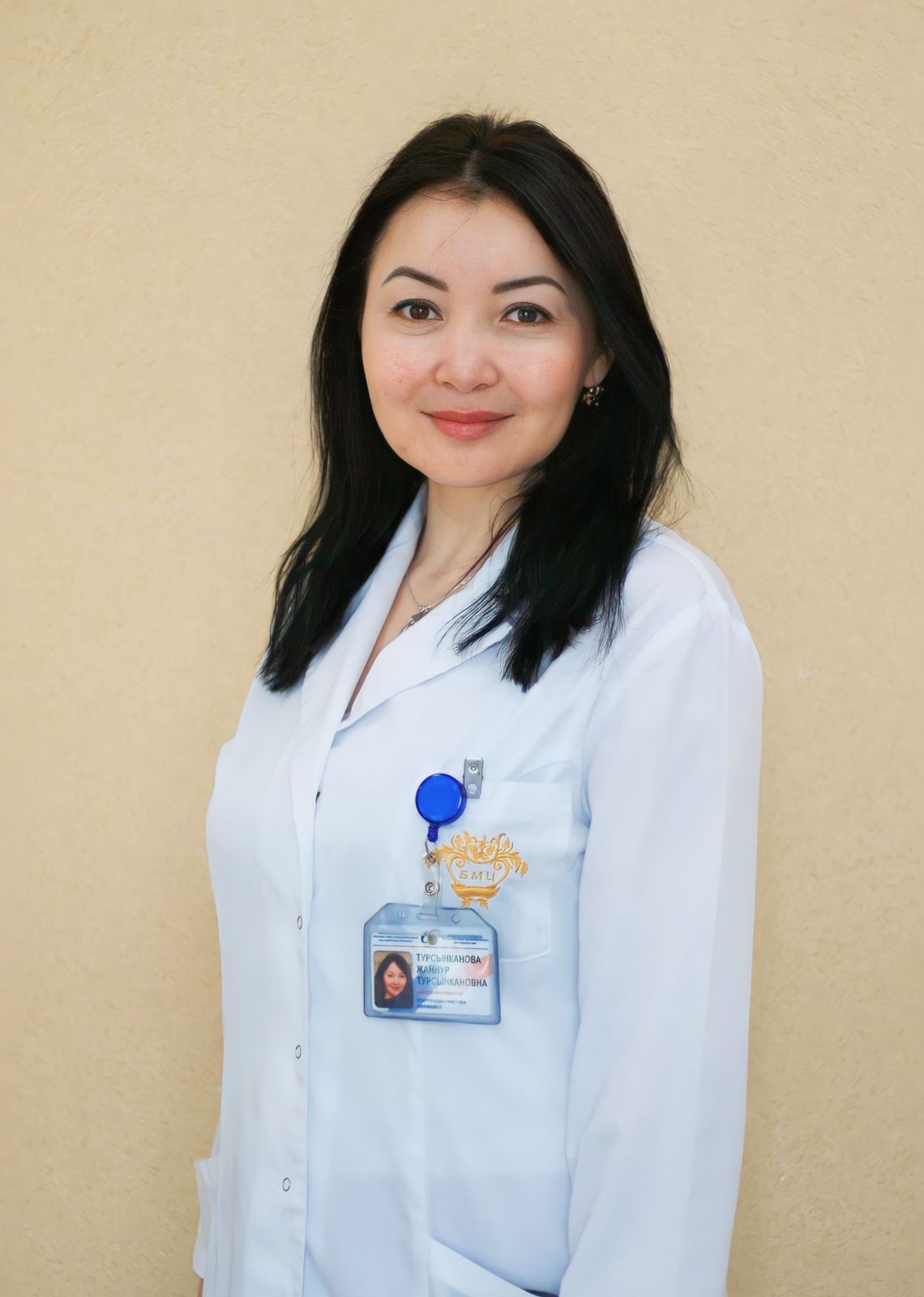 |
Zhannur Tursynkanova Radiologist The first category Work experience: 12 years |
 |
Almagambetov Daniyar Beketovich Radiologist The first category Work experience: 12 years |

