Functional Diagnostics
About the Department
The Department conducts more than 60 types of research, covering the cardiovascular, respiratory systems, neurophysiological research methods. The assessment of the functional state of blood vessels and the heart is carried out on expert-class devices (Vivid e9, Cardiosoft treadmill test system). The Department has a “Sleep Laboratory”that conducts modern methods of diagnosis, evaluation and treatment of sleep disorders with polysomnography, computer pulse oximetry (detection of chronic hypoxemia, sleep apnea) with the selection of treatment (auto – CPAP therapy).
- computer spirometry on automated devices
- spirography with functional tests
- stimulation electroneuromyography
- stress EchoCG
- daily blood pressure monitoring
- transcranial duplex scanning of the brain base (TCCS)
- treadmill test
- holter daily ECG monitoring (12-channel)
- holter daily ECG monitoring (3-channel)
- color duplex scanning of upper limb arteries
- color duplex scanning of lower limb arteries
- color duplex scanning of upper extremity vein
- color duplex scanning of lower extremity veins
- color duplex scanning of ocular arteries and veins
- color duplex scanning of kidney vessels
- color duplex scanning of extracranial brachiocephalic artery vessels
- color duplex scanning of the central retinal arteries
- color duplex scanning of the abdominal aorta and visceral branches
- CDS of arteriovenous fistulas with determination of volumetric blood flow
- CDS of the internal thoracic artery
- transesophageal echocardiography
- ECG (electrocardiography)
- ECG (electrocardiography) with load
- needle electroneuromyography
- electroencephalographic tests (photo,phonostimulation, hyperventilation)
- electroencephalography with computer processing
- EchoCG (echocardiography)
- color duplex scanning of liver vessels
- color duplex scanning of extracranial vessels veins brachiocephalic
- polysomnography
- advanced respiratory monitoring
- electroneuromyography of the upper extremities
- electroneuromyography of the lower extremities
- polysomnography with the selection of CPAP auto mask
- selection of auto CPAP therapy
- respiratory monitoring
- body composition analysis
- selection of BPAPtherapy
- computer spirometry on automated devices
- registration of an EEG attack in the in-patient department
- registration of an EEG attack in the in-patient department in 12 hours
- determination of the diffusion capacity of the lungs with spirography
- ergospirometry
- cardio sonographic examination with bubble contrast (bubble test)
- ECG (electrocardiography) recording
- ECG (electrocardiography) interpretation
- color duplex scanning of testicular veins (CDS of testicular veins)
- EchoCG (echocardiography) with Doppler sonography
- transcranial duplex scanning of the brain vessels with embolus detection (TCDS bubble test)
- nighttime EEG sleep monitoring
- daily EEG sleep monitoring
- EEG sleep monitoring 3-6 hours
- EEG sleep monitoring 6 hours – daytime
- EEG sleep monitoring1. 5 - 3 hours.
Сolor duplex scanning(triplex) of blood vessels of upper, lower limb, blood vessels of the head and neck (CDUS, TCCS), blood vessels, liver, kidneys, abdominal aorta and its branches, transthoracic and transesophageal echocardiography, treadmill stress test, computerized spirometry with bronchodilation tests, daily monitoring of ECG (12 and 3-channel), daily monitoring of blood pressure, electrocardiography, electroencephalography, EEG videomonitoring, stimulation electroneuromyography of upper, lower limbs, computer pulsoximetry, polysomnography, CPAP therapy, the diffusion capacity of the lungs.
The Functional Diagnostics the Hospital has a gas analysis module Power Cube Diffusion by Ganshorn Shiller Group.
This device is designed to determine the diffusion capacity of the lungs by a single inhalation with a breath delay. The results of the DLcO study allow us to judge the ability of the lungs to perform their main function – the transfer of oxygen from atmospheric air to the blood.
Advantages
- Тhe diffusion test is the earliest and most sensitive method of diagnosing disorders of the pulmonary parenchyma structure than spirometry.
- During the study, the patient inhales a gas mixture (helium, carbon monoxide and synthetic air), holds his/her breath for 10 seconds, during which the CO diffuses into the blood, and then makes a forced exhalation. The number of CO passed into the blood is determined and measured in alveolar gas to and at the end of the breath hold, the diffusion capacity of the lungs - the amount of carbon monoxide coming through aero-hematic barrier for 1 min. is calculated.
- Single-use antibacterial filter is used for each study, designed to minimize the risk of air pollution and cross-infection during testing.
- before performing the procedure, the doctor who sends the patient for the examination shall give detailed explanations to the patient about the nature and purpose of the study.
- examination is carried out in the morning, on an empty stomach, for outpatient patients after 15-20 minutes of rest after arriving at the examination room.
- medications that may affect the outcome of the examination are cancelled: bronchodilators are canceled in accordance with their pharmacokinetics: short-acting beta-2 agonists and combined drugs, including short-acting beta - 2 agonists, 6 hours before the examination, long - acting beta-2 agonists-12 hours before examination, prolonged theophylline-24 hours before examination.
- if it is impossible to cancel the medication, the examination shall be conducted against the background of its administration, but when evaluating the results of the respiratory function, it is necessary to take into account the effect of the medication and reflect it in the conclusion.
- chronic diseases of the respiratory system.
- determination of the presence and severity of obstructive and restrictive ventilation disorders.
- with high accuracy, the level of damage to the bronchial tree (large, medium, small bronchi) can be determined.
- in patients with restrictive and obstructive diseases.
- for the diagnosis of emphysema or fibrosis of the pulmonary parenchyma.
- detection of preclinical interstitial lung diseases,
- diffuse fibrosing alveolitis (Hamman-Rich syndrome), pneumoconiosis (silicosis, asbestos, berylliosis), pathological processes leading to a decrease in the gas exchange surface (acute and chronic inflammatory processes), toxic lung lesions.
- development of interstitial edema, development of interstitial fibrosis, sclerotic changes in the parenchyma of the lungs and vascular walls.
- Severe forms of bronchial asthma
- Patients with motor neuroses
- Open forms of pulmonary tuberculosis
- Uncontrolled arterial hypertension>200 mmHg
- Acute myocardial infarction
- Allorhythmia, paroxysmal tachycardia, atrial fibrillation, AV blockades of the 2nd degree
- Unstable angina
- Pregnancy and lactation
Doctors of the Department
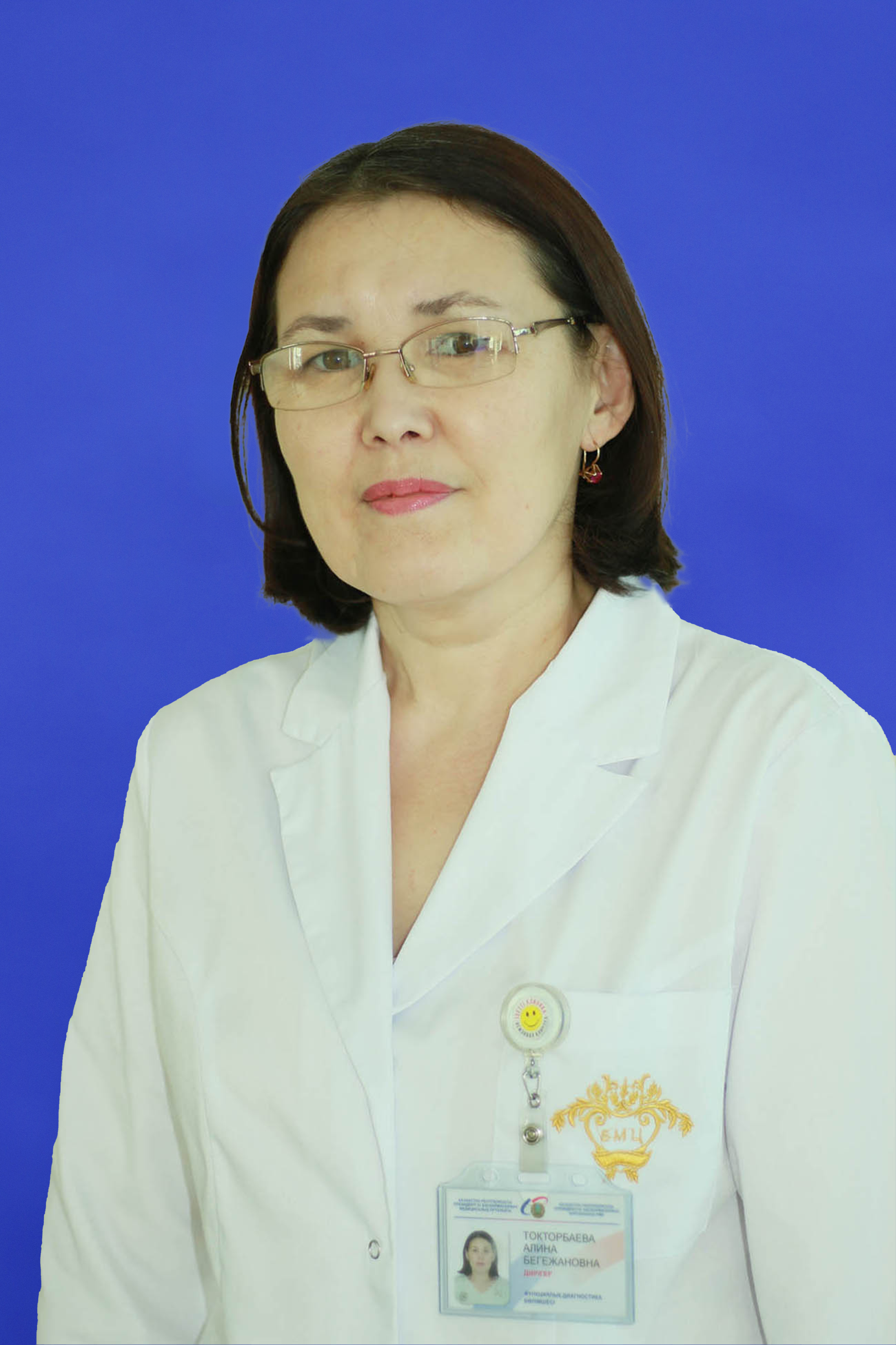 Alina Toktorbayeva
The Highest Category
Work experience: 21 years
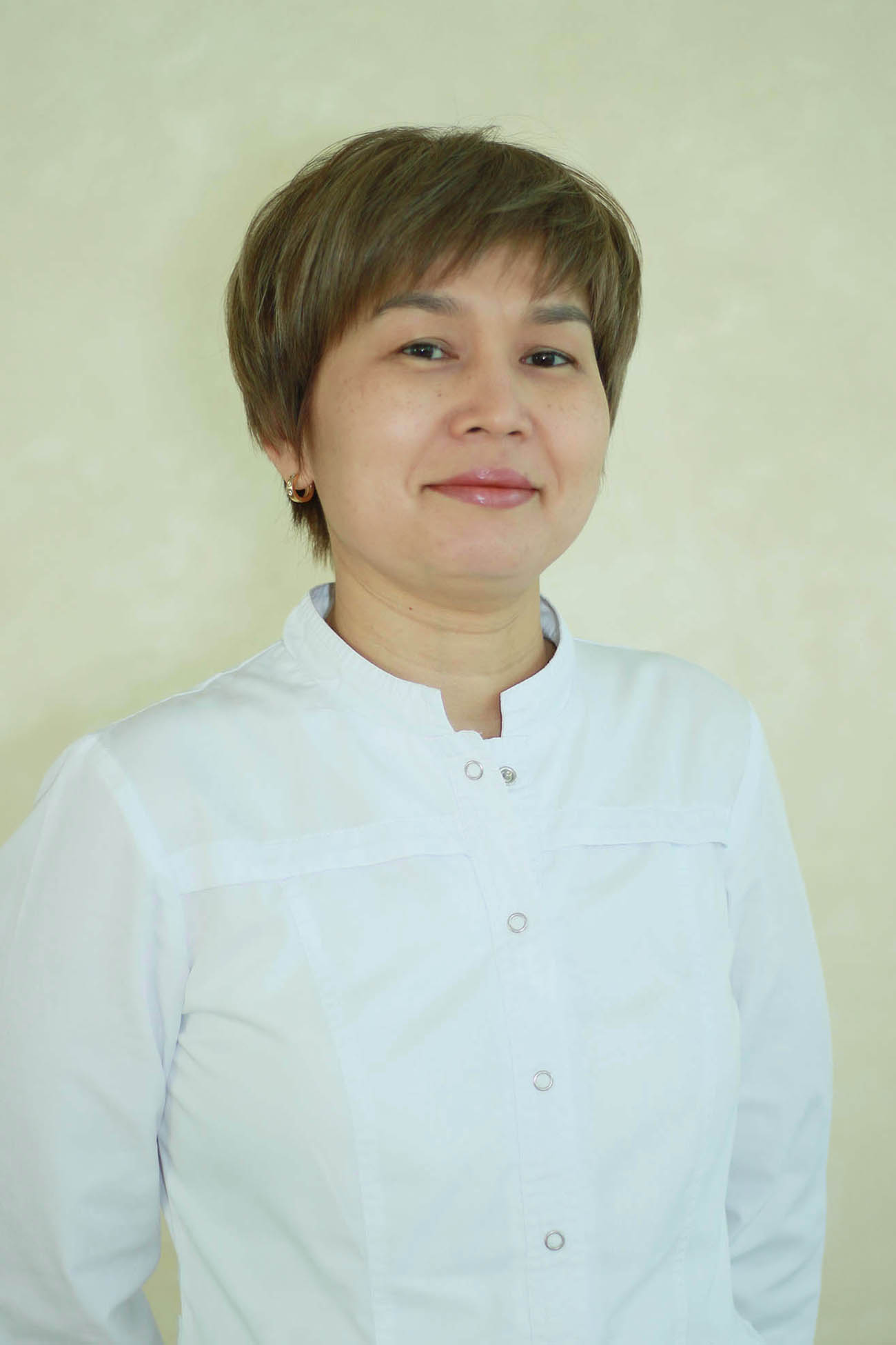 Zaure Seidakhmetovna Baidauletova
 Functional Diagnostics Doctor (EchoCG)
The Highest Category
Work experience: 23 years
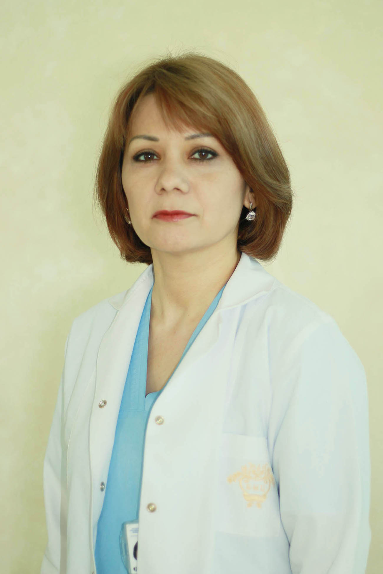 Lyubov Valeriyevna Dushnyak
 Functional Diagnostics Doctor (EchoCG)
Candidate of Medical Sciences
The Highest Category
Work experience: 20 years
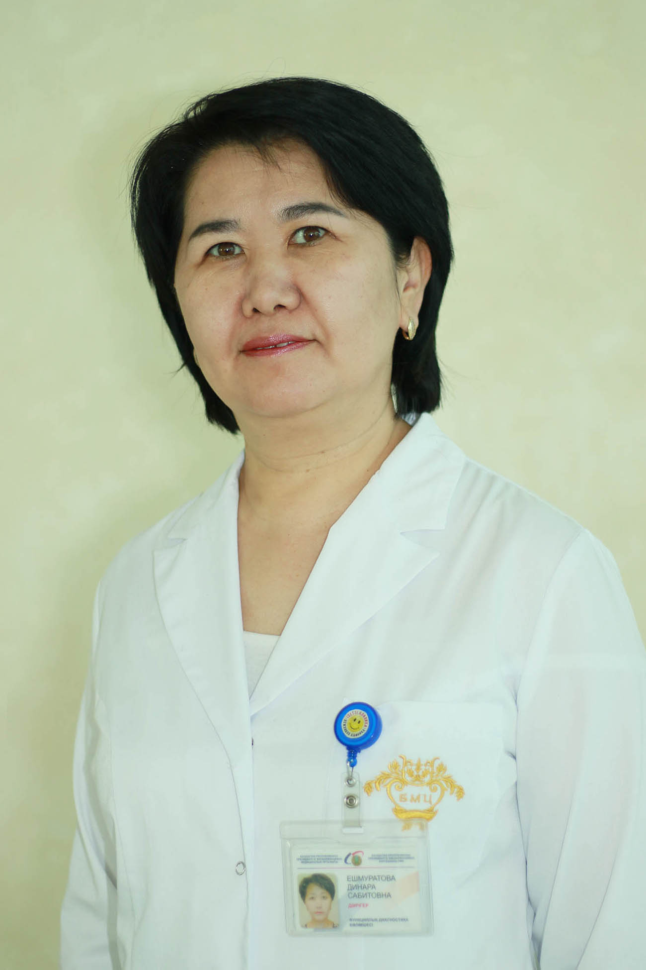 Dinara Sabitovna Yeshmuratova
 Functional Diagnostics Doctor (EchoCG)
The Highest Category
Work experience: 22 years
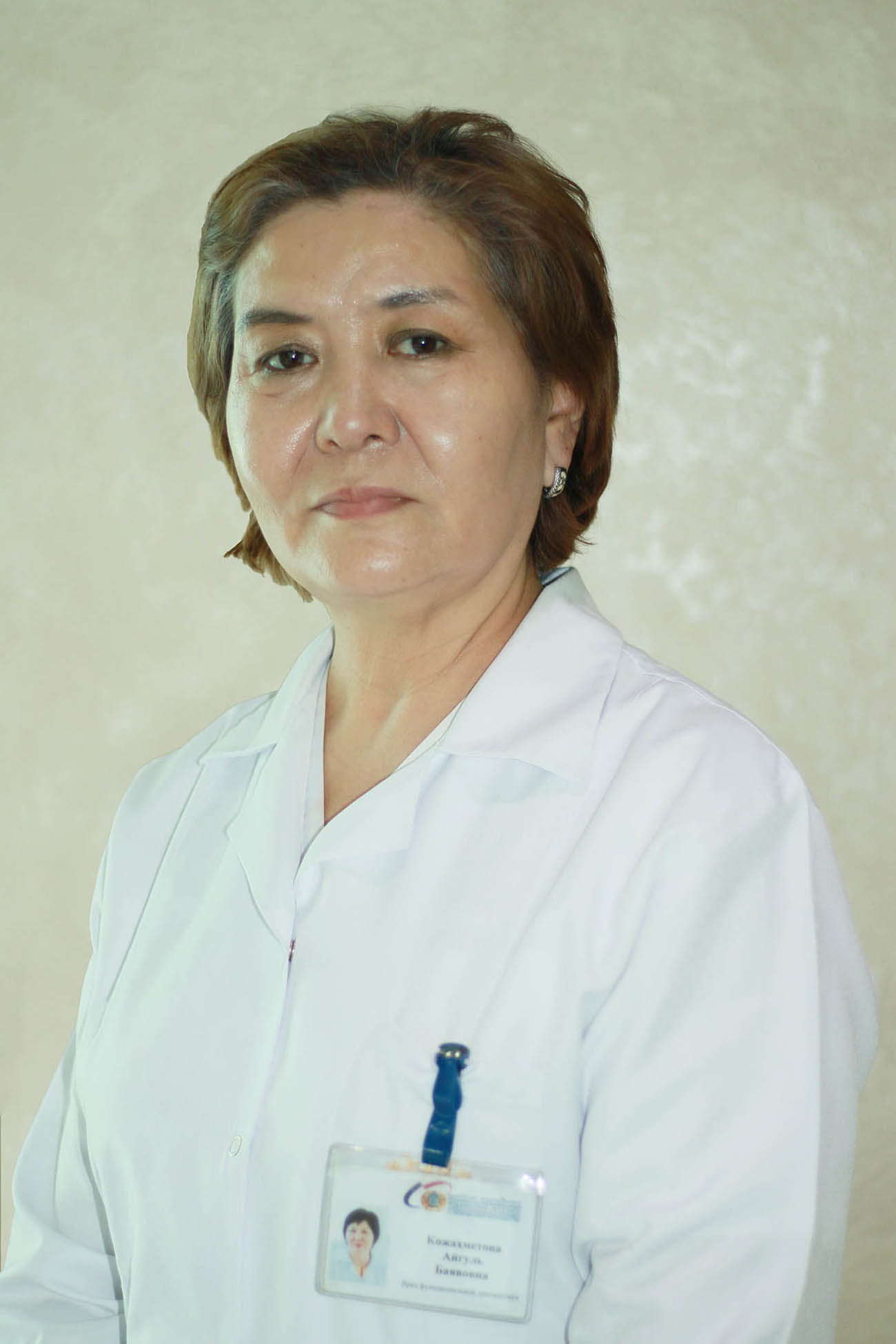 Aigul Bayalovna Kozhakhmetova
 Functional Diagnostics Doctor (EchoCG)
The Highest Category
Work experience: 29 years
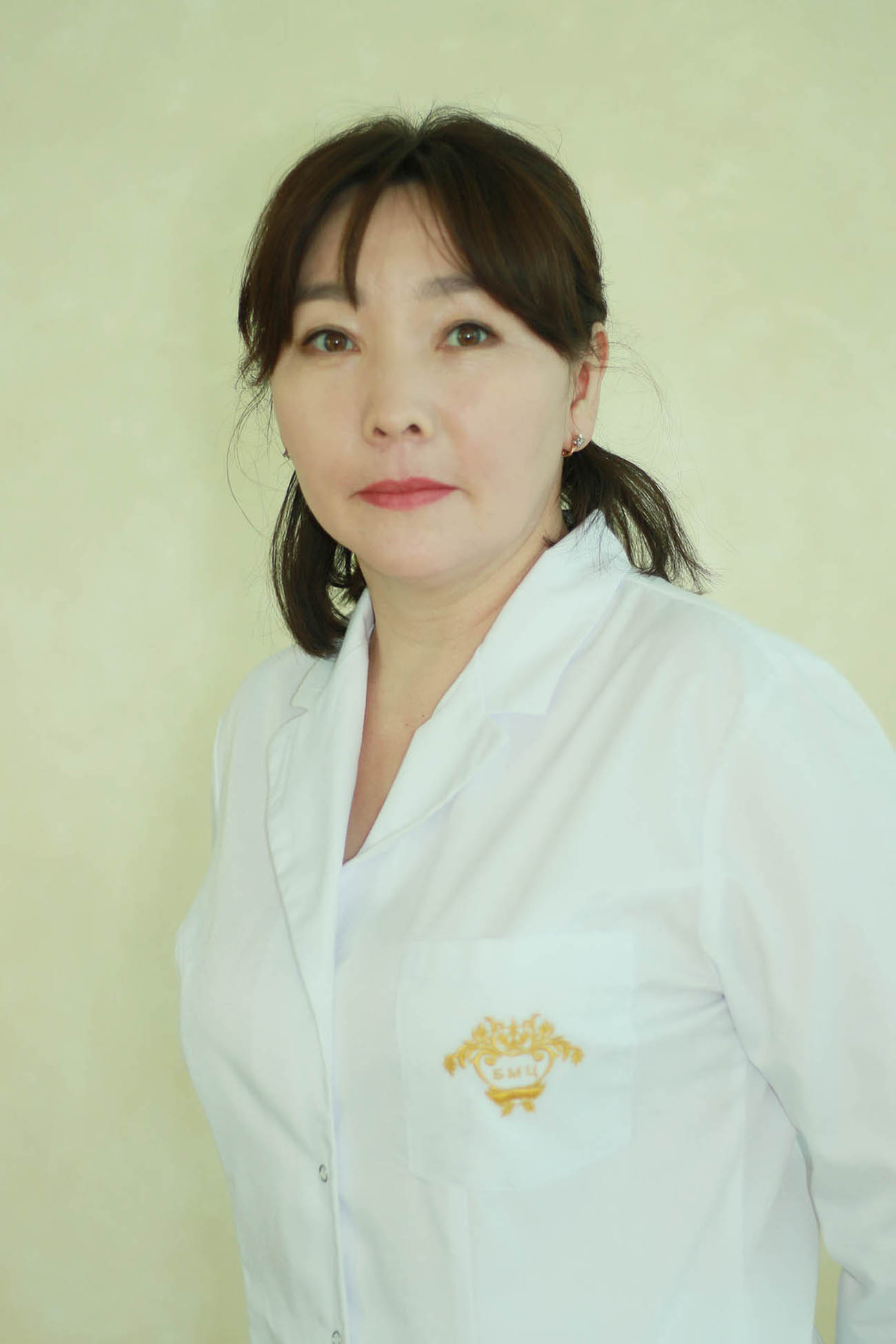 Bayan Khairzhanovna Kulmanova
 Ultrasound Diagnostics Doctor
The Highest Category
Work experience: 29 years
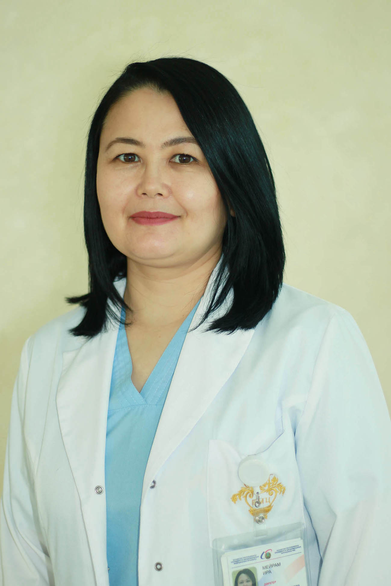 Ira Meiram
 Functional Diagnostics Doctor
The Second Category
Work experience: 10 years
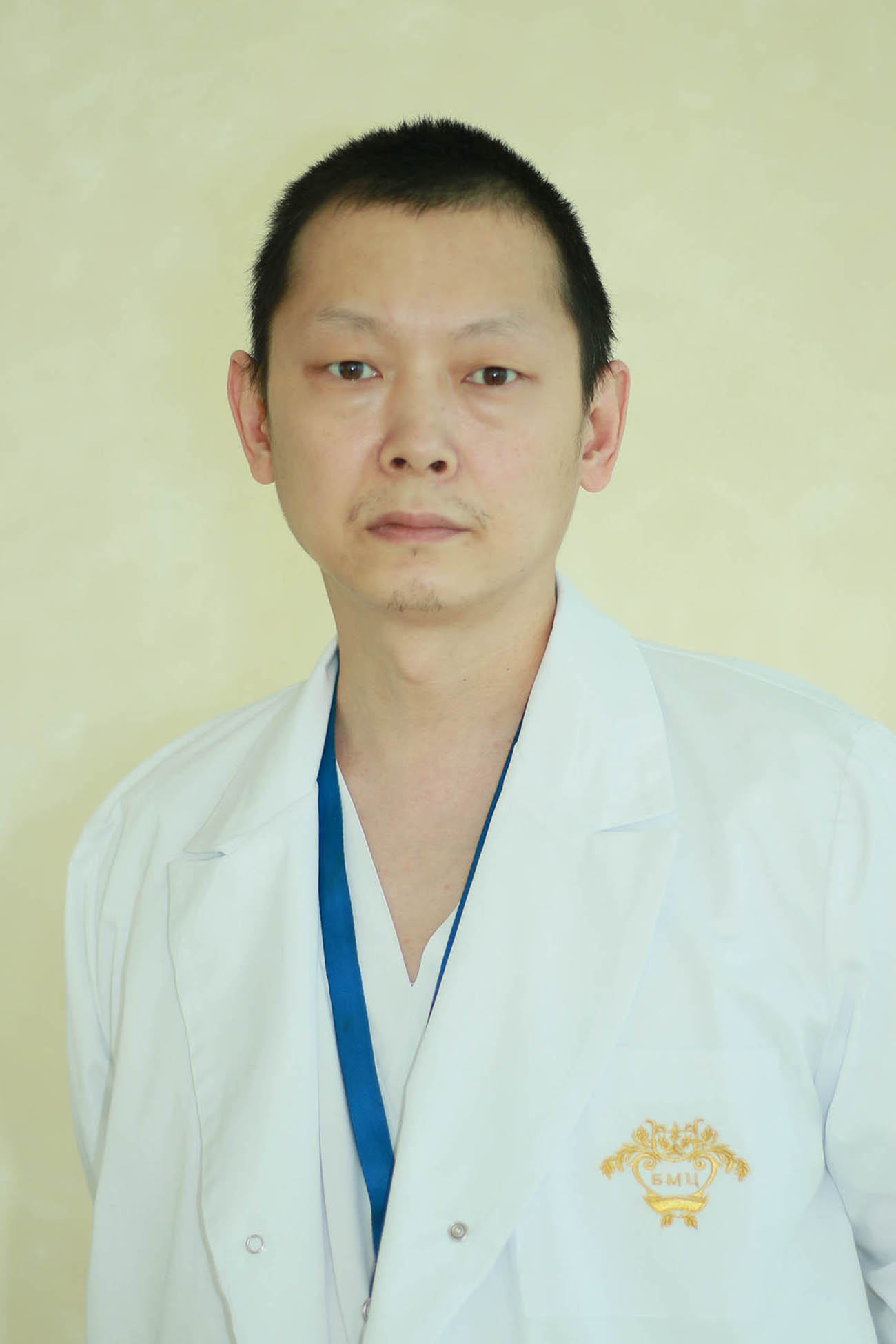 Anatoliy Eduardovitch Min
 Functional Diagnostics Doctor (EchoCG)
The Second Category
Work experience: 20 years
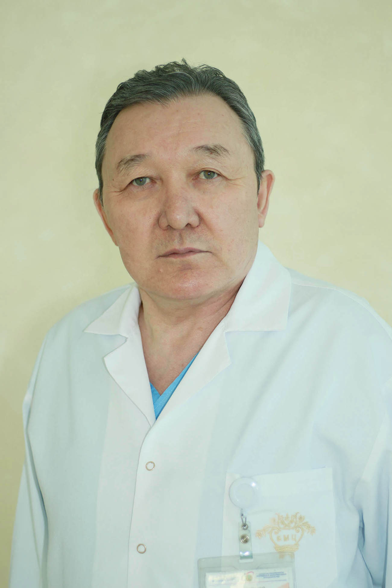 Kayirzhan Toltayevitch Mukanov
 Functional Diagnostics Doctor (EchoCG)
Candidate of Medical Sciences
The Highest Category
Work experience: 38 years
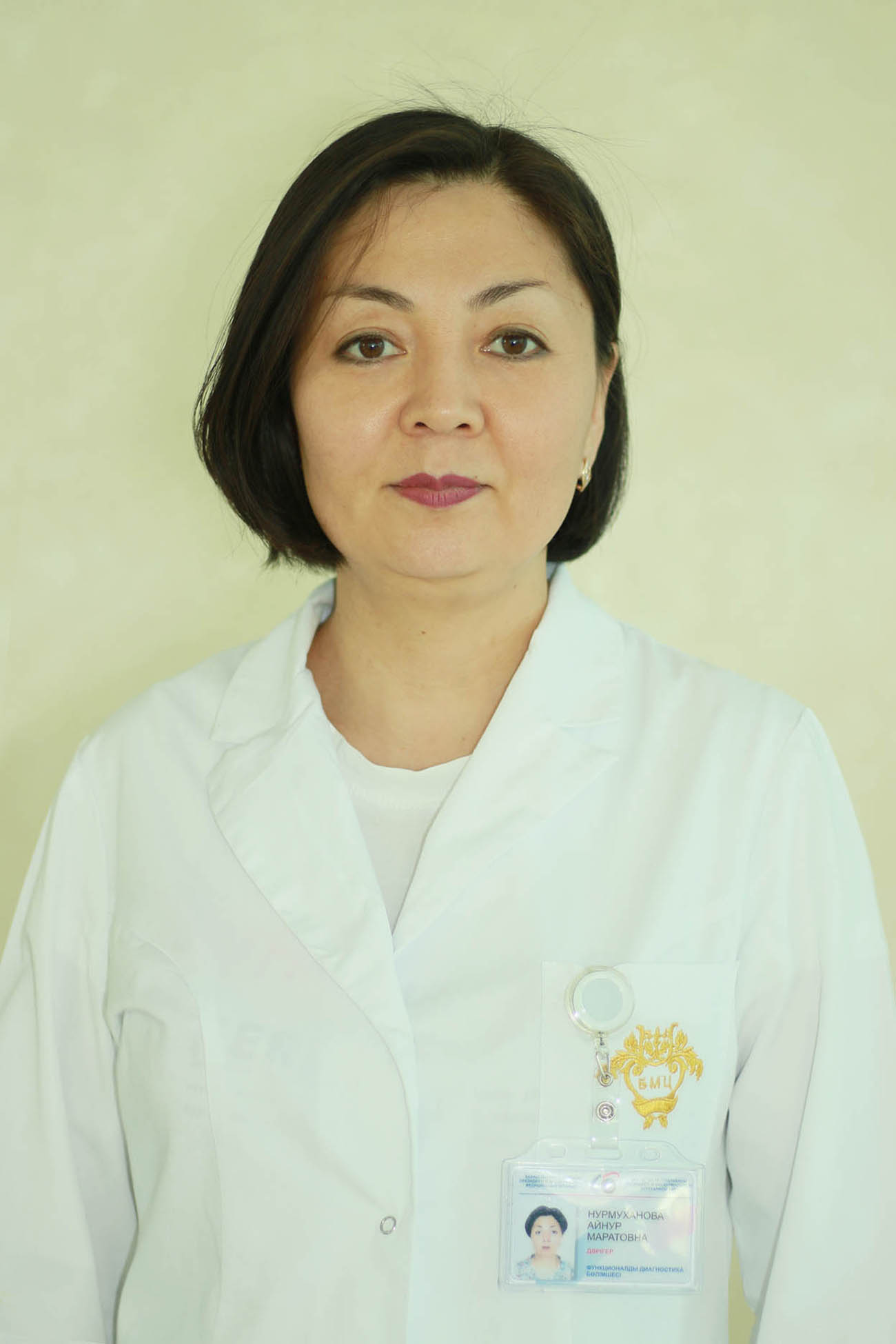 Ainur Maratovna Nurmukhanova
 Functional Diagnostics Doctor (EchoCG)
The Highest Category
Work experience: 16 years
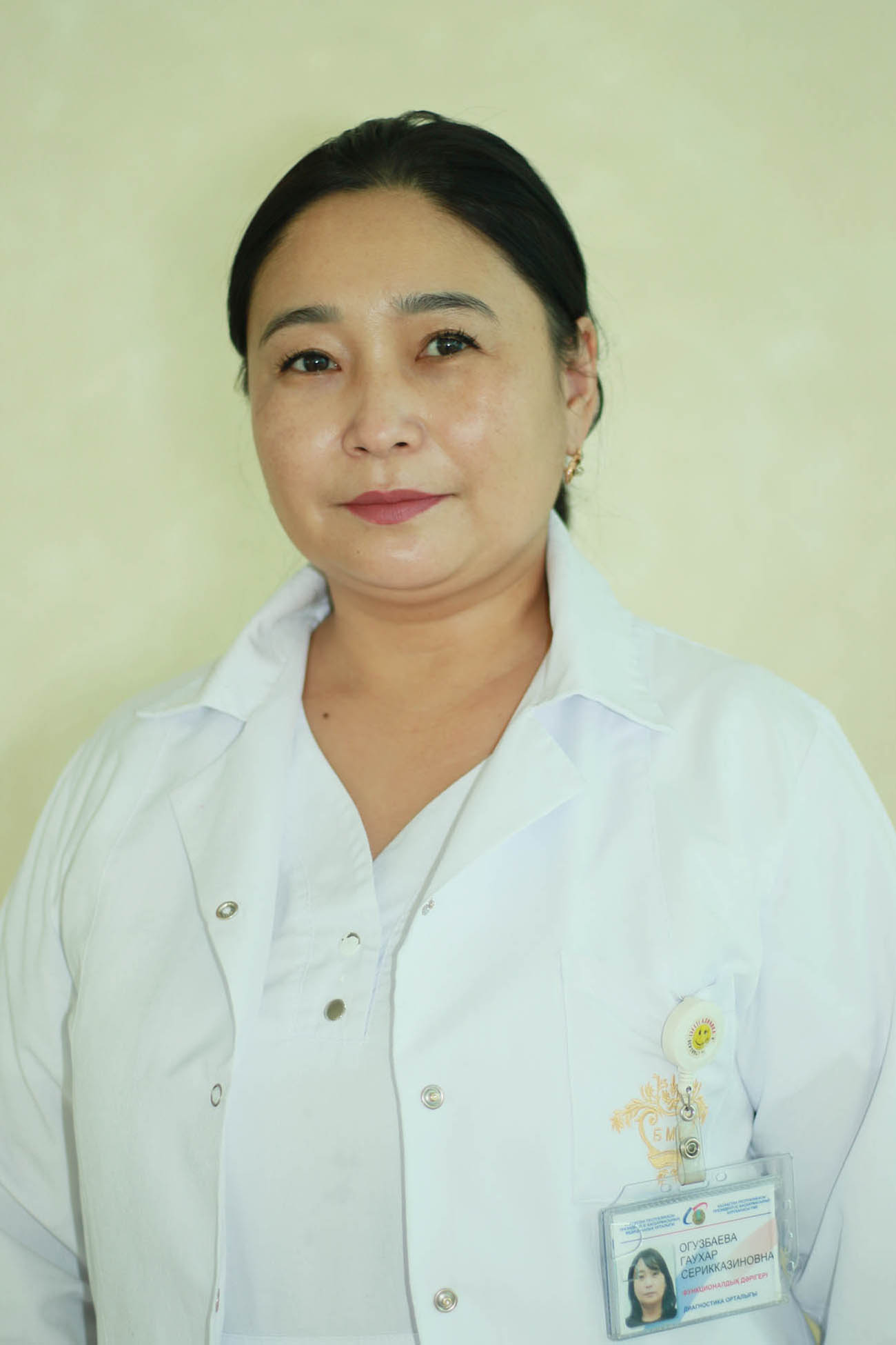 Gaukhar Serikkazinovna Oguzbayeva
 Functional Diagnostics Doctor
The Highest Category
Work experience: 27 years
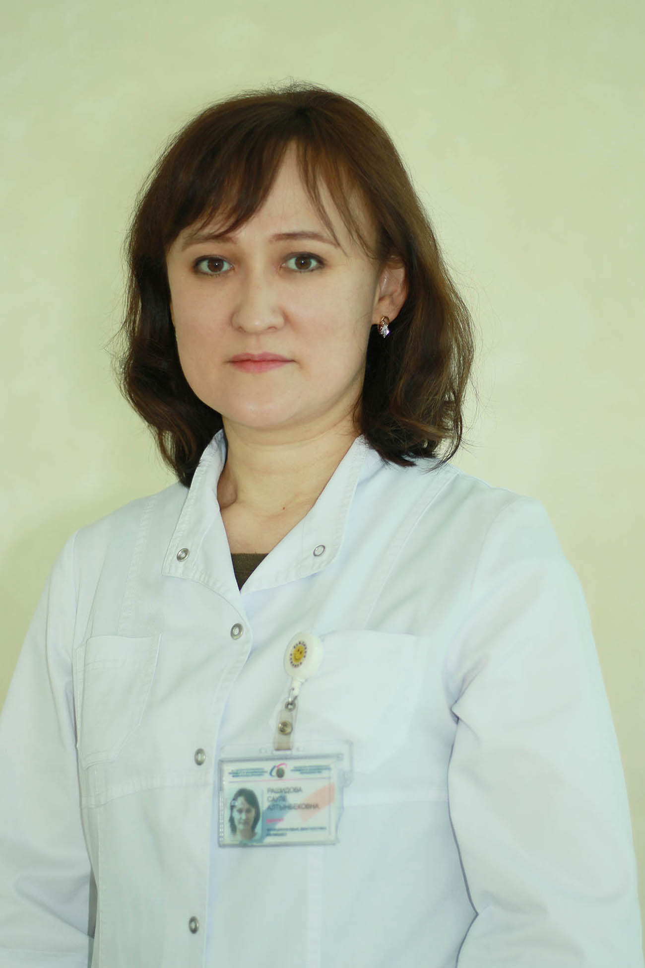 Saule Altynbekovna Rashidova
 Functional Diagnostics Doctor
Work experience: 16 years
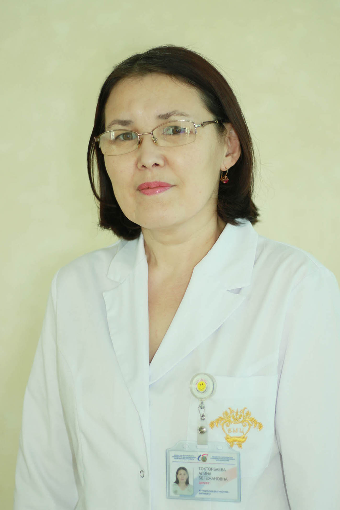 Alina Begezhanovna Toktorbayeva
 Functional Diagnostics Doctor (EchoCG)
The Highest Category
Work experience: 21 years
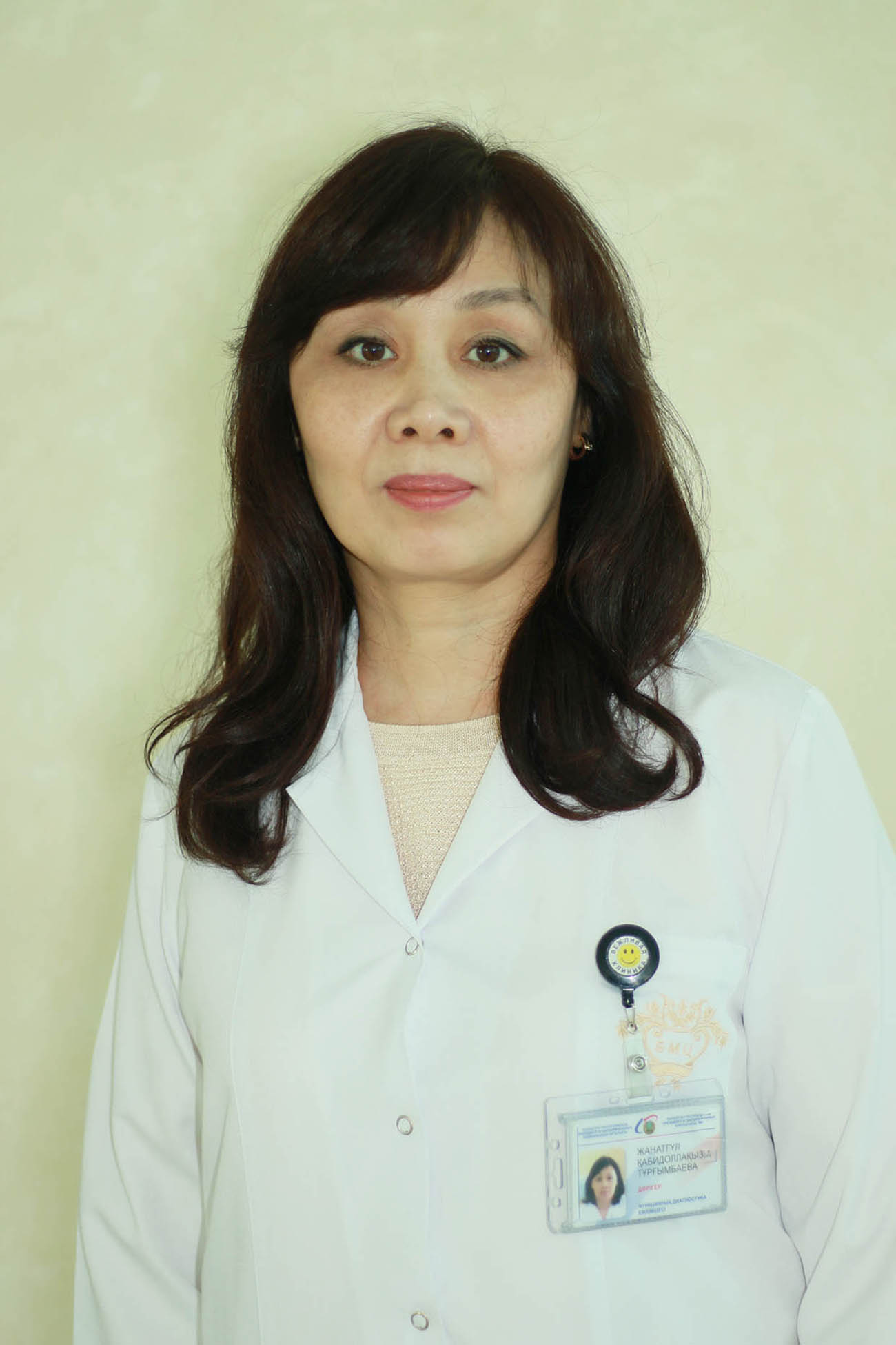 Zhanatgul Kabidullinovna Turgumbayeva
 Functional Diagnostics Doctor (EchoCG)
The Highest Category
Work experience: 29 years
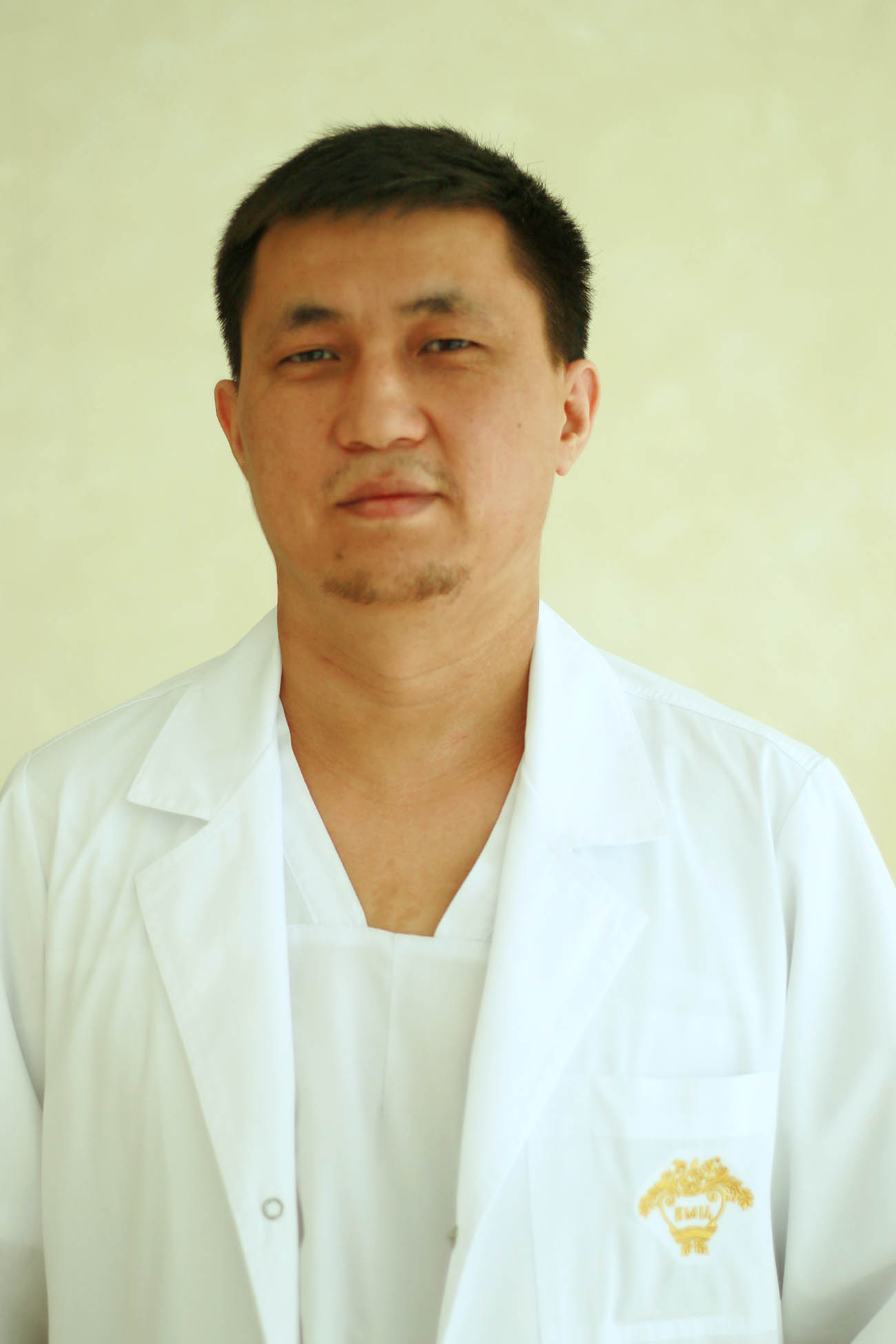 Zhasulan Yergalievitch Utebekov
 Functional Diagnostics Doctor (EchoCG)
The Second Category
Work experience: 10 years
 Olga Vladimirovna Ageyeva
The Highest Category
Work experience: 27 years

